Metabolism, Toxicity, and Deficiency

Background & Metabolism
Selenium was discovered as an essential nutrient in 1957, though it was not discovered what role it played in the body until 1973. The discovery of Se in glutathione peroxidase was the key to understanding its importance in nutrition and health. Glutathione peroxidase, or GSH-Px, is essential for protecting cellular membranes from being destroyed.
Compounds called free radicals are highly reactive molecules, and if left unchecked will destroy cellular membranes. Vitamin E and GSH-Px are two molecules that help prevent this damage. Vitamin E prevents the dangerous molecules (peroxides) from being formed, but even with adequate vitamin E, some peroxides evade destruction. GSH-Px destroys the peroxides before they have a chance to cause membrane damage. GSH-Px concentration and activity is directly related to the selenium status of the animal. Selenium and vitamin E are both antioxidants because they both protect the membranes from oxidative damage. Due to this shared duty, there is a relationship between the compounds, in which one can substitute for the other in a very small way. For instance, more Se is needed when an animal's vitamin E concentrations are low. The sparing effect is an extension of this idea of substitution. Selenium spares vitamin E by:
- preserving pancreas integrity for normal fat digestion, thus normal vitamin E absorption
- reducing the amount of vitamin E needed to maintain lipid membranes via GSH-Px
- aiding in the retention of vitamin E in the blood
Vitamin E spares Se by:
- maintaining body Se in an active form and prevents loss from the body
- preventing destruction of membrane lipids from within the membrane, which inhibits the production of hydroperoxides and decreases the amount of GSH-Px needed
Selenium has also been recently found in another enzyme, 5'-deiodinase. 5'-deiodinase is an enzyme that catalyzes the reaction of the inactive form of thyroxine to the active form. Thyroxine is a very important hormone from the thyroid that helps in regulating body temperature, metabolism, reproduction, circulation, and muscle function. It is known that Se protects the body from heavy metals such as cadmium, mercury, and silver by forming unreactive complexes with them. There are theories that Se may be involved in many other functions in the body, such as-
- a selenoprotein in sperm
- in RNA
- role in prostaglandin synthesis
- role in essential fatty acid metabolism
- required for normal immune response
Elemental selenium (Se (0))can be reacted upon in several ways: it can be reduced to a Se(-2), called selenide, or it can be oxidized to a (+4) state, selenite, or a (+6) state, selenate. Selenium is very similar to sulfur in its chemical properties; it is therefore, not surprising that the main form of organic Se in the body is as selenomethionine and selenocystine. Methionine and cystine are sulfur-containing amino acids, the Se can replace the sulfur because of its chemical similarities to sulfur.
There is not a lot of information on the absorption and pathway of Se from the gastrointestinal tract. It is known that it is absorbed mostly from the upper small intestine; there is no absorption from the stomach, rumen, or abomasum. The amount absorbed depends on the chemical form in which it is ingested. There does not seem to be any feedback loop to reduce the amount of Se absorbed; it has been shown, in rats, that 95% of dietary Se was absorbed regardless if fed deficient or toxic amounts. Absorbed Se travels in the plasma on a protein to its destination tissue. Tissue concentrations vary, the kidneys retain a large amount of Se, along with cardiac and skeletal muscle, and the liver. It is deposited more readily when it is in an organic form. Selenium is readily transferable through the placenta, the mammary barrier, and from hen to egg, so the animal's status will affect offspring and milk concentrations. The primary routes of excretion are through the urine and the feces, exhalation of Se only occurs in cases of toxicity. It has been found that the microorganisms in the rumen may convert Se into insoluble compounds, causing the ruminant animal to absorb less than its monogastric counterpart. It has also been suggested that more Se is absorbed when administered with a high-protein diet, the reasons have not been confirmed.
Distribution/Occurrence
Distribution of selenium is variable throughout the United States. Most of the Eastern and Northwestern areas of the country have low selenium in the soil and in the plants, while in the Midwest, there are adequate amounts of selenium in the forages. For a more detailed map: Map of Selenium Status in US. There are several plant species that accumulate selenium in toxic amounts, and can cause problems in animals which consume them. The most prevalent of these species is the Astragalus genus, which are the locoweeds and milk vetches. For more information on Astragalus:
- Canadian Poisonous Plant Page on Astragalus bisulcatus
- Astragalus Toxicity
- Astragalus (pictures and description)
There are several other species of plants that are characterized as Se accumulators, such as Zylorhiza, woody aster and Oonopsis, goldenweed. All of these plants are more prevalent in the western United States. Highly seleniferous plants are not readily eaten by most animals due to their bitter taste and strong odor, but will be consumed when other forage is sparse.
Feed Analyses
FEED |
Se CONTENT (mg/kg) |
FEED |
Se CONTENT (mg/kg) |
|
Alfalfa |
0.32 - 0.37 |
Barley grain |
0.11 - 0.22 |
|
Brewer's Grain |
0.70 |
Corn (distiller's grain) |
0.48 |
|
Corn (gluten meal) |
1.11 |
Corn (grain) |
0.08 |
|
Cottonseed |
10.0 |
Fish meal |
1.4 - 2.4 |
|
Flax seed |
0.9 |
Oats (grain) |
0.26 |
|
Oats (hay) |
0.17 |
Oats (silage) |
0.01 |
|
Rape seed |
1.05 |
Sorghum silage |
0.21 |
|
Soybean seed |
0.11 |
Sunflower meal |
2.13 |
|
Wheat (soft, winter grain) |
0.05 |
Wheat (hard, winter grain) |
0.45 |
|
Whey (dehydr.) |
0.06 |
Yeast |
0.98 - 1.08 |
|
-This data was taken from the NRC Nutrient Requirements for the different species.
There is only one mineral supplement that contains selenium, and that is selenite.
Toxicity
The toxicity and deficiency sections are general information and contain diseases that affect most species; for species specific diseases, see that species' section.
Selenium toxicity is a serious threat to livestock in the western United States. There is more data on the toxicity of Se in cattle and sheep, but selenosis affects all livestock. There are two general types of toxicity, acute and chronic.
ACUTE TOXICITY
Acute poisoning is caused by the consumption, usually in a single feeding, of a sufficient quantity of highly seleniferous plants, which produce severe symptoms. Usually, death occurs within a few hours. Cattle and sheep are the most likely species to be affected, but also horses, goats, and swine. Studies have shown that possibly as little as 3 mg/kg body weight is the minimum lethal dose in cattle, for horses it may be 3.3 mg/kg BW, and for swine, 1.2 mg/kg BW caused death in 5 days. Symptoms include: abnormal movement, dark watery diarrhea, elevated temperature, weak and rapid pulse, labored respiration, bloating and abdominal pain, mucous membranes are pale and blue, and pupils are dilated. There is no known treatment to reverse the effects of the poisoning, and oftentimes the animal dies before a diagnosis can be made.
CHRONIC TOXICITY
There are two different types of chronic poisoning dependent on the chemical form of the ingested selenium. "Blind staggers" occurs when animals ingest water-soluble selenium compounds naturally found in accumulator plants. Toxicity from eating plants or grain with protein-bound, insoluble selenium is called "alkali disease."
Blind staggers normally occurs in cattle and sheep feeding on seleniferous plants. Symptoms manifest in three stages:
- wandering, stumbling over objects, anorexia, visual impairment
- increase in the severity of the first stage, front legs seem unable to support animal
- blindness, paralysis of tongue and swallowing mechanism, rapid and labored respiration, salivation, and low temperature
 The animal will die within a few hours from the onset of the third stage.
The action of the toxicity has been documented to delay between stages.
The first and second stages may be unnoticeable, and then weeks later,
the animal may show signs of the third stage and die. In sheep, it is more
difficult to diagnose because the stages are not as well defined as in
cattle. Toxic amounts of Se can also cause birth defects in offspring from
dams fed such levels.
The animal will die within a few hours from the onset of the third stage.
The action of the toxicity has been documented to delay between stages.
The first and second stages may be unnoticeable, and then weeks later,
the animal may show signs of the third stage and die. In sheep, it is more
difficult to diagnose because the stages are not as well defined as in
cattle. Toxic amounts of Se can also cause birth defects in offspring from
dams fed such levels.
Alkali disease is more chronic than blind staggers, often taking years
to manifest itself. It is caused by feeding on plants and grain that have
protein-bound, insoluble selenium. This disease can affect all livestock,
but it is detected mostly in cattle and horses. General symptoms include:
lack of vitality, anemia, emaciation, stiffness of joints, lameness, rough
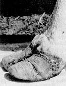 coat, loss of long hair, and hoof sloughing and deformities. Hoof deformities
are a classic sign of selenium and can cause lameness and severe pain for
the animal; food and water must be provided to the animal, for it may be
hesitant to walk.
coat, loss of long hair, and hoof sloughing and deformities. Hoof deformities
are a classic sign of selenium and can cause lameness and severe pain for
the animal; food and water must be provided to the animal, for it may be
hesitant to walk.
PREVENTION
The most effective way of preventing selenosis is to remove the animals from the seleniferous area. Treating the soil with sulfates, thus changing the S:Se ratio, can sometimes depress Se uptake by accumulator plants. Results from studies have shown that feeding a higher protein diet may reduce the toxicity of Se; animals fed the same amount of toxic selenium but fed a higher protein diet lived for a few more days than those animals fed a low protein diet. Dilution of high Se feeds with low Se feeds in a mixed ration will help to prevent toxicity. Recognition of seleniferous plants, proper land management, and grazing control are all necessary to completely prevent selenosis.
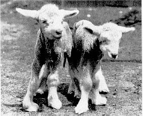
Deficiency
Deficiency of Se is much more common in the eastern United States where the soil content of Se is low. There are many different diseases that affect the different species, all of which will be addressed in the species sections. There is one disease that is consistent in all livestock species, and that is Nutritional Muscular Dystrophy, or White Muscle Disease (WMD). Nutritional muscular dystrophy is caused by the deficiency of Se and/or vitamin E and S-containing amino acids. The disease is characterized by degeneration of the skeletal muscles, causing stiff gaits, and other problems.
PREVENTION
The most effective way of preventing Se deficiency is to supplement concentrates fed to animals with commercially available supplements to a level of about 0.1-0.3 mg/kg Se. Sodium selenite is the most common commercial supplement; calcium selenite is also useful, and is less hazardous than sodium selenite. For animals that also eat forage, it would be possible to supplement low Se diets with high Se plants and grains. There are also Se-supplemented salt licks available, and may be the most viable option to farmers. Fertilizer containing Se has not been shown to significantly increase the Se content of the plants from that soil. In areas of known Se deficiency, it is often practice to give calves and second trimester pregnant cows intramuscular injections of Se and vitamin E to prevent disease.
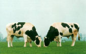
Cattle
The nutrient requirement for Se for dairy cattle has been set at 0.3 mg/kg, while the requirement for beef cattle is 0.2 mg/kg. Requirements are higher when legumes are fed, S intake is high, vitamin E intake is low, and when diets contain heavy metals. Diets with high amounts of unsaturated fatty acids will increase the requirement for Se. Effects of Se deficiency in cattle include, white muscle disease, retained placenta, unthriftiness, cystic ovarian disease, and anemia.
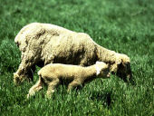
Sheep
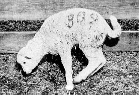 The nutrient requirement for Se for sheep is 0.10 - 0.20 mg/kg. Sheepshare some of the same diseases that cattle get. There is high incidence
of white muscle disease in lambs, and there are two types of WMD that affect
lambs. The first is congenital WMD, where lambs are wither stillborn, or
die within a few days, after physical exertion. The second type is delayed
WMD, and can occur from 1-4 months after birth. These lambs walk with an
unsteady gait and arched back.
Ewes also suffer from infertility and embryonic loss when Se deficient.
There is also a general unthriftiness, i.e., growth depression, decrease
in food consumption, and decrease in wool production.
The nutrient requirement for Se for sheep is 0.10 - 0.20 mg/kg. Sheepshare some of the same diseases that cattle get. There is high incidence
of white muscle disease in lambs, and there are two types of WMD that affect
lambs. The first is congenital WMD, where lambs are wither stillborn, or
die within a few days, after physical exertion. The second type is delayed
WMD, and can occur from 1-4 months after birth. These lambs walk with an
unsteady gait and arched back.
Ewes also suffer from infertility and embryonic loss when Se deficient.
There is also a general unthriftiness, i.e., growth depression, decrease
in food consumption, and decrease in wool production.
Swine
The nutrient requirement for Se for swine is 0.10 - 0.30 mg/kg. Pigs may show a variety of diseases in relation to Se deficiency. Hepatic dietetica is the degeneration of the liver, which can manifest itself acutely in rapidly growing pigs as liver failure, or more sub-acutely as jaundice, edema, and/or cardiomyopathy. Mulberry heart disease occurs in growing pigs that develop severe cardiomyopathy, often coupled with hemorrhages of the cardiac tissue. Similar to the other species, pigs can also develop nutritional muscular dystrophy. Spermatogenesis decreases when Se intake is extremely low. Pigs are also more susceptible to swine dysentery when selenium deficient.
Horses
The nutrient requirement for horses is 0.10 mg/kg. Nutritional muscular dystrophy is the known disease that affects horses with Se deficiency. Similar to sheep, there are three different patterns of NMD that can occur. The first is acute, with death occurring within 24 hours. The foal's tongue may be paralyzed, making in unable to suckle. The second case is more common and is induced by exercise. Older foals are more susceptible to this form; they show an unsteady gait and general muscle weakness, rapid heart rate with arrhythmia, and labored breathing. After a few days, it is difficult to make them stand and they salivate excessively. Mortality from this condition is only about 30-45%. The third condition affects mostly older animals, and is the result of chronic Se deficiency. Affected animals show anorexia, emaciation, generalized muscle weakness, rapid heart rate, and diarrhea.
Poultry
The nutrient requirement for poultry varies with the status of the animal. For young chicks, < 6 weeks, the requirement is 0.15 mg/kg; for the others, the requirement is 0.10 mg/kg. Several conditions affect poultry due to Se deficiency. One is exudative diathesis, which is the accumulation of fluid throughout the body, particularly in the abdomen and feet. This is caused by increased permeability of the capillaries and leakage of fluid from the capillaries. Chicks with this condition are also anemic and are protein deficient. It occurs about 2-4 weeks after hatching and is easily diagnosed due to the edema and the blue-green tint to the skin after progressing to the hemorrhagic stage. Poultry are also affected by nutritional muscular dystrophy. They also suffer from pancreatic atrophy, which has been found to be caused solely by Se deficiency. Atrophy of the pancreas results in a reduction in the amounts of lipase, trypsinogen, and chymotrypsin--all enzymes that aid in digestion of food. Therefore, this leads to extremely reduced growth and feathering. Egg production decreases when hens are Se deficient; there is no evidence that suggests an effect on male reproduction.
References
Barbezat, G. O., C. E. Casey, P. G. Reasbeck, M. F. Robinson, and C. D. Thomson. Selenium IN:Current Topics in Nutrition, vol. 12. Alan R. Liss, Inc., New York, 1984.
Combs, G. F. and S. B. Combs. The Role of Selenium in Nutrition. Academic Press, New York, 1986.
James, L. F., K. E. Panter, H. F. Mayland, M. R. Miller, and D. C. Baker. Selenium Poisoning in Livestock: A Review and Progress. IN:Selenium in Agriculture and the Environment. American Society of Agronomy, Inc., Madison, Wisconsin, 1989.
McDowell, L. R. Minerals in Animal and Human Nutrition. Academic Press, New York, 1992.
National Research Council. The Nutrient Requirements of Sheep.. National Academy Press, Washington DC, 1968.
National Research Council. The Nutrient Requirements of Beef Cattle.. National Academy Press, Washington DC, 1984.
National Research Council. The Nutrient Requirements of Swine.. National Academy Press, Washington DC, 1988.
National Research Council. The Nutrient Requirements of Dairy Cattle.. National Academy Press, Washington DC, 1989.
Rechcigl, M. Jr. CRC Handbook Series in Nutrition and Food--Section E: Nutritional Disorders, vol. 1. CRC Press, West Palm Beach, FL, 1978.
Rosenfeld, I. and O. A. Beath. Selenium: Geobotany, Biochemistry, Toxicity, and Nutrition.. Academic Press, New York, 1964.
Underwood, E. J. The Mineral Nutrition of Livestock.. Commonwealth Agricultural Bureaux, England, 1981.
Background & Metabolism] [Distribution/Occurrence] [Feed Analyses] [Toxicity] [Deficiency] [Cattle] [Sheep] [Swine] [Horses] [Poultry] [References] [Return to list of toxicants]